Tuesday, 20 November 2007


The creation of new glucose is GLUCONEOGENESIS

A rise in blood glucose concentration:
1) receptors pick up that there is high glucose in the blood
2) the beta cells in the islets of Langerhans secrete insulin directly into the blood stream.
3) insulin increases the permeability of of cell membranes to glucose , so more glucose is absorbed.
4) Insulin also activates the condensation of glucose to glycogen in glycogenesis
5) insulin also increases the rate of respiration of glucose.
6) blood glucose decreases


A decrease in blood glucose concentration:
1) alpha cells of the islets of Langerhans secrete glucagon into the blood stream.
2) this activates the hydrolysis of stored glycogen to glucose . this is called glycogenolysis
3) blood glucose increases

Pancreas:
key organ in controlling blood glucse levels
cells are known as the islets of langerhaans, are endocrine function.
Alpha cells secrete: glucagon
beta cells secret: insulin

these hormones are secreted straight into the blood capillaries
they are antagonistic

Insulin is a hormone that decreases the level of glucose in the blood. Two effects of insulin are cells are more permeable to glucose, speeds up acquisition. it also activates enzymes stimulation the conversion of glucose to glycogen.
Its main target organs are the Pancreas, Liver, muscles.

Glucagon hormone that increase blood sugar, main effect is that it activates enzymes which are responsible for converting glycogen into glucose (glycogenolysis)

the brain only respires glucose

glycogenesis: the synthesis of glycogen from glucose under the influence of insulin

glycogenolysis: the breakdown of insoluble glycogen under the influence of glucagon to form glucose.
gluconeogenesis: the creation of glucose from non carb sources e.g. lipids/ fatty acids or protein.

test for glucose:
diabur strips, yellow= no glucose,
progressively darkgreen-blue= glucose present

Benedicts test:(less sophisticated)
not specific to glucose just for redcuing sugars
after heating for 3 mins a blue solution means no glucose.
a brick red means glucose

Tuesday, 13 November 2007

The kidney

The kidney
The kidneys are organs that filter waste products out of the blood and excrete then e.g. urea along with water as urine.
Urea is made by the liver. Excess amino acids can’t be stored therefore the liver breaks them down to urea.(ornithine cycle)
OSMOREGULATION: The homeostatic mechanism of the active regulation of the osmotic pressure of bodily fluids to maintain the homeostasis of the body’s level of water, minerals and salts in the blood.
NITROGENOUS EXCRETION: excretion of nitrogen in urine.

The kidney is involved with the homeostatic functions of regulating the chemical properties of the blood. (Water, solvent concentration and pH)


When the blood passes through the kidney nephrons liquid is filtered out of the blood, carrying solutes with it, including urea. Useful nutrients are selectively reabsorbed and the wasted products are removed in urine.
There are two main stages of kidney function:

1) ULTRAFILTRATION
This occurs in the barrier between the blood and the filtrate in the Bowman’s capsule. In the Bowman’s capsule there is a dense network of capillaries called the glomerulus, blood flows into the capillaries.
A glomerulus is enclosed in the sac. Fluids from blood in the glomerulus are collected in the Bowman's capsule (i.e., glomerular filtrate) and further processed along the nephron to form urine. This process is known as ultrafiltration.
A high pressure caused by the blood moving into the capillaries via the afferent wide arteriole and moving out through the narrow efferent arteriole. The high pressure forces small molecules such as
water, glucose, amino acids, sodium chloride and urea through the filter, from the blood in the glomerular capsule across the basement membrane of the Bowman's capsule and into the nephron. This type of high pressure filtration is ultrafiltration.
Blood is forced against the capillary endothelium which has pores in it which allow the blood plasma to reach the basement membrane and small molecules are squeezed
about but large ones like protein and blood cells stay in.

2) Selective reabsorption (proximal tubule)
Selective reabsorption takes place in the proximal convoluted tubule (PCT) of the kidney. Blood leaving the Bowman’s capsule has a low hydrostatic pressure and so flows slowly. It has a high concentration as these could not pass out before. It therefore has a low water potential and this enables it to gain water from the proximal tubule. It is the process by which certain substances that are required by the body (such as glucose, amino acids, vitamins and water) but have been filtered out of the blood during ultrafiltration are reabsorbed. As only certain substances are reabsorbed, it is known as selective reabsorption.

substances secreted into the distal convoluted tubule are: h+ ions and potassium ions.
substances actively reabsorbed from the proximal convoluted tubule are sodium ions and glucose.
the kidney tubule has adaptation such as lots of mitochondria and a brush border for atp for the active reabsorption of molecules and a large surface area for absorption.
overview: sodium ions are actively pumped out, glucose reabsorbed, amino acids reabsorbed and chloride reabsorbed, 50 % urea reabsorbed

ADH makes the collecting duct more permeable to water. So more water is taken back into the body by osmosis. more water is taken back into the blood stream and less is lost in urine. It is secreted by the posterior pituitary gland. Osmoreceptors detect changes in the hypothalamus.

Urine: composition varies due to diet and climate.


Thursday, 8 November 2007

Unbalanced diet and diseases

Under nutrition

A lack of protein in the diet is called protein-calorie malnutrition. This is commonly found in the developing world in poorer countries.
Conditions caused by lack of protein:

Marasmus- malnutrition caused by a diet low in protein and energy/carbohydrates. Also caused by early weaning off breast milk. Suffers are grossly underweight, they have thin arms and legs, no body fat and muscle is wasting away.

Kwashiorkor- malnutrition caused by low protein only. usually occurs in slightly later age than marasmus. Suffers have oedema (swelling) in the feet and lower legs, pale thin hair, muscle wasting and a moon face.

Anorexia nervosa and bulimia nervosa are malnutrition due to psychological issues.

Two examples of Under-Nutrition are Anorexia nervosa and Bulimia nervosa.
These are examples of poor dieting, resulting in a lack of essential nutrients. They are eating disorders and the under-nutrition is self induced.
Anorexia nervosa usually starts in the teenage years, mainly affecting women and girls but boys can be affected.
The disease is characterised by a list of symptoms:
1. Body weight is maintained at 15% below that expected for a persons height.
2.Self induced weight loss, from forced vomiting, taking laxatives, avoiding fatty foods and excessive exercise.
3. Fear of putting on weight, obsessed with the size of their body.
4. Pursue a very low ideal weight.
5. Can cause hormonal differences and women can stop having periods.

Normally seen in the western world, from social and biological factors, peer pressure, media and stress can cause Anorexia nervosa.

6. Delayed development of puberty.
7. Feel bloated after a small meal.
8. loose interest of socializing with friends
9. They feel tired
10. Constipation
11. Feeling cold
12. Low blood pressure

Can lead to:
1.Chronic illness
2. Death
3. Osteoporosis
4. Damage to heart, kidneys, liver and brain.
5. Difficulties in concentration
6. Mental health problems

Bulimia nervosa

This is another eating disorder which involves an irresistible urge to 'binge' eat on food, followed immediately by self induced vomiting. laxatives etc are used as well to prevent weight gain.
Vomiting can lead to loss of potassium, chloride and hydrogen ions.

symptoms:
1. Dental erosion
2. Sore throat
3. Swollen salivary glands
4. Fatigue
5. Constipation
6. loss of voice
7. dry skin, brittle nails
8. Anemia
9. Infertility
10. Liver failure


people who suffer from Bulimia nervosa do not show the symptoms of extreme weight loss.
The person will be of normal or higher weight, the person is less likely to drop significant amounts of weight.
This is less to do with food but more with psychological issues.

Vitamin A
retinol is found in dairy products and fish liver oils. It is needed to keep lining tissues healthy.(epithelium). Deficiency leads to night blindness.

Vitamin C
Ascorbic acid is found in citrus fruits, green vegetables etc. it is needed for hydroxylase to work. It maintains collagen fibres in connective tissue. Deficiency is scurvy as the connective tissue weakens.

Calcium
Found in dairy products, bread and flour. calcium is absorbed through the small intestines by carrier proteins. The carrier can only be made with vitamin D. lack of vitamin D can lead to calcium deficiency. This leads to rickets, muscle spasms and osteomalacia.

Iron
found in both meat and plants. its needed for haemoglobin in red blood cells. deficiency leads to anaemia

Sunday, 14 October 2007

Metabolic Pathways

Metabolism
This is the sum total of all the reactions occurring in the cells.
reactions include the breakdown of glucose to CO2 and H2O.
These reaction take place in a series of steps e.g reactants----- intermediates-----products
Each reaction in a pathways is catalysed by an enzyme.
The products can act as an inhibitor of an enzyme so it can control its own production.

Catabolism: the breakdown of complex molecules into smaller ones e.g. break down of glucose
This usually creates ATP
Anabolism: the synthesis of complex molecules from simple ones. e.g. joining amino acids together.
this usually uses ATP

The cell has two ways of controlling metabolic reaction:
1) compartmentalisation e.g. reactions take place in specific areas of the cell
2) enzymes, which can be inhibited by their products.

OXIDOREDUCTASES catalyse oxidation and reduction
HYDROLASES catalyse hydrolysis reactions e.g. breaking molecules byt he addition of water. used in : digestion etc


cellular respiration

All living organisms require energy for three major reasons:


1) for mechanical work e.g. muscle contraction

2) the active transport of ions across cell membranes

3) the synthesis of macromolecules


This 'free' energy is obtained from the environment.


Cellular respiration is the oxidation of food substances to obtain 'free' energy. Free energy can be used to drive reaction that require an input of free energy, such as active transport.

It occurs as a series of linked, enzymes catalysed reactions.

1) Gylcolysis

2) Krebs cycle

3) oxidative phosphorylation


Atp- Adenosine Triphosphate
this is the most important product of cellular respiration.
Used at an immediate source of free energy rather than a long term store. Its rate of production is high and usually used with 1 minute of synthesis.
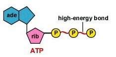


adenosine-ribose(5 carbon sugar)-3 inorganic phosphate groups


This is important because the phosphoanhydride bonds yield a high amount of free energy when hydrolysed.Atp can be hydrolysed to ADP (adenosine diphosphate) and Pi and hydrogen.


ATP+ H2O---> ADP + P(i) + H(+)

deltaG= -30 kj mol-1


This is an exergonic reaction as it yields free energy.


ATP is quickly resynthesised from the products when food substances are oxidised in chemotropic organisms or due to light trapping on photosynthetic organisms.

Electron Carries


Many reactions in metabolic pathways require the removal of electrons or hydrogen atoms in the oxidation of substrates.


The electrons are transferred to a group of substances known as electron carriers (aka coenzymes or hydrogen carriers)


The reduced form of these carries transfer their electrons to oxygen by a chain of electron carriers in the inner mitochondrial membrane. ATP is formed from ADP and Pi as a result.


NAD(+) = Nicotinamide adenine dinucleotide.


when a substrate is oxidised NAD accepts a hydrogen ion and 2 electrons. the reduced form is NADH. only 1 hydrogen atom is passed on


NAD(+) + 2H(+) + 2e(-) ----> NADH + H(+)

overview:


1) Intake of Food substances


2) substrate oxidised (oxidation removes hydrogen and electrons)

3)NAD(+)---reduced by addition of hydrogen and electrons to NADH

4)These reduced forms transfer their electrons to oxygen in the inner mitochondrial membrane

5) ATP is produced as a result of the energy released in the process below. the energy is used to generate another process in the cell to make atp.


(2NADH + 2H+ + O2 ==> 2NAD+ + 2H2O) dont have to learn!


Glycolysis

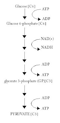
This occurs in the cytoplasm of cells and is a series of enzyme-catalysed reactions in which each molecule of glucose is turned into 2 molecules of pyruvate.


Pyruvate: a compound containing 3 carbon atoms and links glycolysis to the next reaction.


glucose + ATP ------(hexokinase)-------> glucose 6-phosphate + ADP + H(+)


Phosporylation of glucose:


1)prevents glucose from leaving the cell, as the membrane is impermeable to sugar phosphates.


2) it makes glucose more reactive


summary:


Two 3-carbon compounds of Pyruvate are made from each molecule of glucose.


2 molecules of ATP used but 4 produced in glycolysis.


NET PRODUCTION :TWO MOLECUlES OF ATP


Pyruvate then passes into a mitochondrion and then the krebs cycle occurs.


Aerobic Respiration(oxygen present)

The krebs cycle

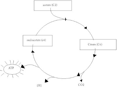
Pyruvate created in glycolysis passes into the matrix of a mitochondrion. The link reaction now occurs, where Pyruvate is turned into acetate and added to coenzynme A, forming ACETYL COENZYME A. Also NADH is formed and CO2.

Pyruvate + NAD(+) + CoA ---------> Acetyl CoA + NADH + CO2


4 carbon compound combines with with two carbon acetyl to form acetyl CoA, this forms a 6 carbon compound citrate.


2 atoms are lost in each turn of the cycle. These are lost as CO2.


NAD reduced and FAD

Hydrogen is lost


summary:

Two carbon atoms enter the carbon cycle and two carbon are lost as carbon dioxide

one molecule of ATP is formed

4 pairs of hydrogen are removed

3 NAD's are reduced and 1 FAD is reduced

The reduced electron carriers are reoxidised in the electron transport chain.

Oxidative Phosphorylation


this is the process by which atp is made. ATP is formed when electrons are transferred from NADH or FADH2 to oxygen.NADH oxidation produces 3 ATP, FADH2 oxidation produces 2 ATP.


Each [H] atom splits into [H+] and [e-]


The transfer of electrons to oxygen through electron carriers leads to [H+] being pumped from the matrix into the inter membrane space. when they flow back into the matrix the free energy made avaliable is used to make ATP.


Electrons are transfered from NADH to oxygen through a series of large protein complexes.


Oxygen acts as the final electron recpetor and is reduced to form the final product which is , water.

ATP YIELD: 36 molecules from one glucose molecule.

Anerobic respiration:

the electron transport chain cannot function without oxygen. Respiring anerobically poses a problem as they need to reoxidise electron carriers.

used during vigorous physical exertion.

Pyruvate is reduced to Lactate and NAD so glycolysis can continue.

lactate accumulates in the muscles and after exercise lactate is oxidised back to pyruvate.

In yeast it ends up with ethanol and CO2 produced, the yield of ATP is two molecules from one molecule of glucose.

--